A five-volt ultrasound patch advances the engineering of continuous monitoring
A Beijing team has demonstrated a wearable ultrasound array driven at five volts peak. Its November 2 paper in Microsystems & Nanoengineering points toward smaller electronics for continuous cardiovascular monitoring.
Published November 4, 2026Analysis / Frontier Tech / Human Augmentation
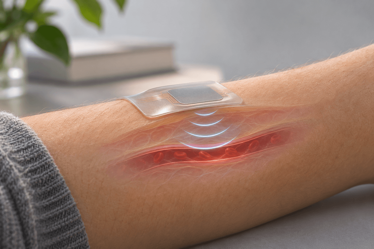
Tong Jin, Hang Gao and colleagues at the Institute of Microelectronics of the Chinese Academy of Sciences worked with Tsinghua University and the Chinese PLA General Hospital. Differently sized vibrating membranes merge their acoustic responses, producing 81.7 percent bandwidth and artery images within four centimeters. The primary research paper
Ultrasound forms an image from returning sound waves. The time an echo takes to arrive helps locate a structure; its strength provides further information. Repeating those measurements can show movement, including changes in organs and blood vessels. The U.S. Food and Drug Administration describes ultrasound as a real-time imaging method with applications ranging from blood-flow assessment to echocardiography. That ability to measure movement is central to the interest in a patch that stays in place over time. FDA's explanation of ultrasound imaging
Converting an examination tool into a continuously worn sensor is a systems problem. The transducer must maintain contact, electronics must produce and record the signals, and software must turn the measurements into useful information. A low drive voltage can help the electronics design. Overall energy use also depends on how often the array transmits, how signals are processed and how data are communicated. Those quantities determine the battery and thermal requirements of an eventual wearable product.
In six volunteers, blood-pressure measurements after cuff calibration averaged a 2.2-millimeter-of-mercury deviation from a clinical reference. The nine-hour monitoring trace came from one subject. These are prototype feasibility results; regulatory approval of the reference monitor applies to that monitor. Study methods and results
The patch joins several approaches to wearable ultrasound already under investigation. In a 2023 Nature study, Hongjie Hu, Hao Huang, Sheng Xu and collaborators described a wearable cardiac imager with a deep-learning method for extracting left-ventricular volume from continuous recordings. That work illustrates another layer of the problem: following anatomy and converting image sequences into measures such as cardiac output and ejection fraction. The transducer, the measurement target and the interpretation method all shape what a wearable system can do. The earlier cardiac-imaging study
Another project led by Muyang Lin and Xu, announced by the University of California San Diego in May 2023, combined a wearable ultrasound probe with wearable control electronics and wireless monitoring. It provides useful context for the current engineering race: one research program may concentrate on the acoustic component, another on integration and operation while a person moves. Comparing them by the particular function demonstrated makes their contributions easier to understand. UC San Diego's account of the integrated system
Fabrication used six-inch silicon-on-insulator wafers. Development goals include wafer-uniformity and reliability measurements, stretchable interconnections and improved performance during movement. These connect the research to the practical demands of manufacturing consistency and comfortable use. Fabrication and future work
A five-volt transducer demonstration does not establish a clinically validated, fully integrated monitoring product. The next useful evidence would cover a broader participant population, varied daily movement, long-term signal stability, complete-system power use and the clinical decisions the measurements can support. Each test answers a different question, from whether the patch stays coupled to the skin to whether a derived measurement remains reliable across people and conditions.
The potential value of continuous ultrasound is the time dimension: a record of how physiology changes during activity, rest or a clinical event. Turning that record into useful care will require agreement among sensor engineering, clinical validation and interpretation. The new low-voltage array advances one component of that effort and gives researchers a concrete design to develop alongside the rest of the system.