A temporary ultrasound implant could help doctors watch an injured spinal cord
After surgery to relieve pressure on an injured spinal cord, doctors still need to know what is happening near the damage. Closing the wound makes that harder. Johns Hopkins researchers are testing a temporary ultrasound implant intended to keep that area visible during recovery.
Published November 8, 2026Briefs / Frontier Tech / Experimental Systems
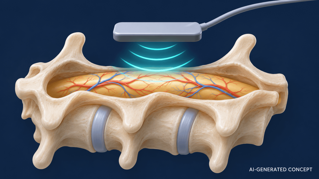
After surgery to relieve pressure on an injured spinal cord, doctors still need to know what is happening near the damage. Closing the wound makes that harder. Johns Hopkins researchers are testing a temporary ultrasound implant intended to keep that area visible during recovery. Research data and abstract
The device, called MUSIC, is designed to sit over the spinal cord and provide images and information about blood movement. Johns Hopkins reported its first animal proof of concept on November 4, alongside publication in Science Advances. The work is still preclinical; it has not established that patients recover better. Johns Hopkins report
The distinction matters because the project has two ambitions: observing the injured cord and eventually using focused ultrasound to influence local blood circulation. The researchers demonstrated imaging and tracked local blood-motion signals. Their exploratory ultrasound intervention was tested in healthy pigs, rather than proving that it restores circulation in injured patients. Authors’ data archive
“Timing is critical for recovery,” Nicholas Theodore, who developed the device with biomedical engineer Amir Manbachi, said in the university’s announcement. The intended benefit is practical: give care teams information near the injury while reducing the need to move a vulnerable patient for repeated imaging. Johns Hopkins report
What the measurements actually show
The authors’ public data documentation is unusually useful for understanding the limits. Its focused-ultrasound analysis covers 30 repeated sessions across three healthy-pig experiments. The measured quantity was Doppler-derived blood-flow velocity: an estimate of how fast blood was moving. It was not a direct measurement of blood volume delivered to tissue or evidence of therapeutic benefit. Data documentation
That leaves an important clinical question. A faster-moving signal alone cannot tell doctors whether damaged tissue receives enough oxygen, whether an intervention protects nerve cells or whether someone will regain movement. A useful monitoring device would need to produce information that helps clinicians make better decisions, and that benefit would have to be tested.
A long development path remains
The project grew from a $13.48 million DARPA award announced in October 2020. At the time, Johns Hopkins described an ambitious five-year effort toward human use. The new animal study illustrates why a development target should be read as a goal rather than a promised clinical arrival date. Original project announcement
Johns Hopkins says MUSIC received FDA Breakthrough Device designation in 2023. That designation offers closer interaction with regulators and prioritized review; it is not marketing authorization. Devices in the program still have to meet the FDA’s safety and effectiveness requirements. Johns Hopkins report · FDA explanation